RADIOLOGY: DEVELOPING CORE IMAGING SKILLS
| Course Overview |
|
Radiography provides veterinary practices with a vital diagnostic tool. The value of that tool, however, is directly related to the quality of the images obtained. Poor technique can result in missing a foreign body, bone cancer or a fracture. Congestion in the lungs - is it fluid or mucous? Masses? What about the radiograph of that swollen abdomen? Is the radiograph of sufficient diagnostic quality to reveal if it is ascites or an abdominal mass? Besides the risk of missing or misinterpreting a problem, poor radiographs cost the practice money. The cost of retakes in staff time can reduce the income from the procedure to almost nil. On a busy day, repeated takes can greatly increase stress to the team as well as to the patient. The ability to take quality radiographs is a combination of education and skill.
Many team members are given a cursory introduction that does not provide all the needed training. Although quality training requires an investment of both time and money, the benefits to the practice, the patient, the client, and the team greatly outweigh the cost of poor radiographic technique. Radiology: Developing Core Imaging Skills is a 6-week course that provides participants with the training needed to understand the fundamentals of the functioning radiographic systems, as well as safety concerns and exposure monitoring. It is recommended, although not required, that participants work in a practice to complete this course.
|
| Continuing Education Credits |
|
Course meets the requirements for 15 RACE hours of continuing education credit for veterinary technicians in jurisdictions which recognize AAVSB's RACE approval. However, participants should be aware that some boards have limitations on the number of hours accepted in certain categories and/or restrictions on certain methods of delivery.
This course is an interactive online course that meets RACE requirements; program number 57-19150
|
| Course Content |
Radiology: Developing Core Imaging Skills is a 6-week course that begins with an overview of image acquisition, including developing an understanding of cathodes, anodes, focal spots, distance, time, mAs, kVp and general machine use. Positioning aids, film markers and identification, logs and filing protocols, safety and personal protection are covered in depth.
Small animal technique and positioning instructions, along with pictures, help the participant focus on the appropriate method for obtaining the best diagnostic image results for the abdomen, thorax, chest, shoulder, scapula, skull, spine, pelvis, forelimb and hindlimb. Participants will be able to develop technique charts, and make adjustments to optimize the image.
All positioning instructions include pictures with directional targets to show where the x-ray beam should be focused. Avian and exotic techniques and positioning overviews and tips are provided as well. This course also addresses general dental radiography including the anatomy of the dental radiograph machine, dental film and film processing along with small animal dental positioning, including troubleshooting tips and technique usage. Contrast studies and patient considerations as well as an overview of ultrasound rounds out the course.
|
| Course Focus and Learning Objectives |
Radiology: Developing Core Imaging Skills is a course that is designed for any member of the veterinary team wishing to enhance their skills in radiograph acquisition.
Upon completion of this course, the participant will be able to:
- Differentiate between analog, computerized and digital radiography
- Identify artifact and film and implement changes to eliminate
- Describe transducers, image modes and how ultrasound images are produced
- Identify radiation risks and implement mandatory safety measures
- Compare and contrast proper positioning methods to obtain ideal radiographs
- Apply non manual restraint and animal handling, so that the patient does not need to be manually restrained when being radiographed
- Compare and contrast the advantages and disadvantages of various positing aids available
- List positioning guidelines to ensure production of good quality diagnostic radiographs
- Identify normal radiographic anatomy of the abdominal and thoracic cavities
- Properly and safely position a dog or cat for the various common positions, with an emphasis on where to measure, center the beam, where the borders are, and how to properly position, so that the body part is parallel to the film and both are perpendicular to the central ray
|
| Participation Access Parameters |
|
The design of this course allows participants to enroll at any time.
After enrolling, please allow up to 48 hours for course activation.
Each participant will have a personal start and end date that begins upon activation.
|
| Required Materials |
Lavin's Radiography for Veterinary Technicians, 5th Edition Marge Brown
Important Text Information:
It is mandatory that each participant has access to this textbook as the content of the book is not reproduced within the course. It is up to each person to determine the best way to acquire the text. Some will already have the text in their library and, therefore, will not need to purchase another copy. The text is available as a print publication and as an eBook. Where to purchase and in what format is totally up to the participant. The text is not included in the fee for this course.
USA residents: Elsevier texts, both print and eBook, are available via the VetMedTeam Elsevier Text Portal at discounts off regular retail of 25 - 35 %. If you would like to visit the portal to take advantage of the discount please use this link:
VetMedTeam's Elsevier Text Portal
Important: Regardless of which option the participant chooses, access to a copy of the textbook is mandatory. Without the text, the student will not be able to complete the assignments, case studies and examinations. If enrolling close to or beyond the course start date, it is recommended that the text be purchased as an eBook to prevent text acquisition related delays. VetMedTeam has no control over shipping delays and other related problems.
|
| Course Completion Requirements |
This is an instructor led course, with no real time components or lectures. Completion requirements include:
- Instructor Graded Interactive Module Assignments: Designed to help the participant reinforce newly learned material
- Instructor Graded Interactive Case Study Assignments: Designed to help the participant apply the newly learned skills and knowledge though patient and practice based scenarios
- Examinations: All examinations must be submitted with a score of 80% or better
- Survey
|
| Participant Feedback |
|
The entire course was well put together, and I think the case studies were most beneficial to me. I enjoyed looking at and thinking of what was done on the samples and how I could or would improve the image. I have a strong interest in radiography and always strive to be better. I feel that this course has helped me understand how an xray works and have a better appreciation for the diagnostic abilities.
I thoroughly enjoyed viewing rads as our case studies and having to rethink ways that would improve upon the image.
I liked the Technical details (kV and mAs) and also non-manual methods of restrain
Having the Lavin text is a great reference.
|
| Course Instructor |
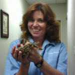 Helen E. Sweeney, DVM Helen E. Sweeney, DVM
Dr Helen Sweeney is the owner of Elma Animal Hospital and Aquatic Veterinary Services of WNY. She has been in private small and exotic animal practice since receiving her DVM degree from the University of Georgia. Dr. Sweeney is a member of several national and regional professional organizations including AVMA, ARAV, AEMV, AAFV, WAVMA, NFVS, WNYVMA, and NYSVMS. She served as the President of the Niagara Frontier Veterinary Society (NFVS), was selected for two terms as Chairman of AVMA's Aquatic Veterinary Medicine
Committee, and currently serves as the past President of the American Association of Fish Veterinarians, the AAFV, which she helped found.
Dr. Sweeney is a frequent speaker at national and international veterinary conferences on pet fish health. She is an instructor for AquaVet® (a summer Aquatic Animal medicine program for veterinary students associated with Cornell University college of Veterinary Medicine). She edited and co-authored 'Fundamentals of Ornamental Fish Health', published by Wiley-Blackwell late 2009. She has authored and co-authored several articles published in peer reviewed journals and texts and is a consultant for veterinary
Information Network. Dr. Sweeney was also featured in 'Fixing Nemo', an article on fish veterinarians published in the New York Times Magazine.
Dr. Sweeney has a special interest in public health and is the Veterinary Medical Consultant for the Buffalo Veterans Affairs Hospital animal research facility.
In her spare time, Dr. Sweeney enjoys photography and gardening. She has two fish ponds in her backyard, her own private oasis. Many other pets complete the 'zoo', including two mixed breed dogs, Phoenix and Gryphon; and 'Spike', a vintage tuxedo cat.
|
| Pricing |
Price: $179.00 USD
This course is eligible for the 10% Multi-course discount. To learn more about the discount please visit the Multi-Course Discount page.
|